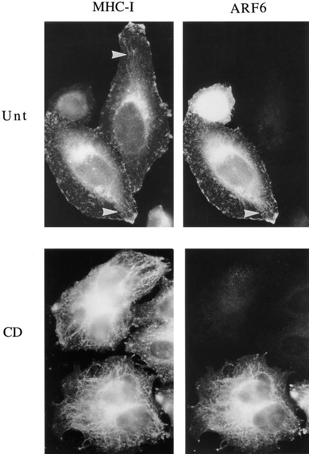
Figure 4.
Endogenous MHC-I localizes to the tubular compartment with or without ARF6 overexpression. HeLa cells were transfected with ARF6 plasmid and then incubated in the absence (Unt) or presence (CD) of 1 μM CD for 30 min at 37°C, and they were fixed and processed for immunofluorescence localization of MHC-I using mouse anti–MHC-I antibodies and for overexpression of ARF6 using antibody to ARF6. Anti–MHC-I was detected with sequential FITC-labeled goat anti–mouse and FITC-labeled donkey anti–goat antibodies, while anti-ARF6 was detected with rhodamine-conjugated donkey anti–rabbit antibody. The tubular structures (arrowheads) radiating from the perinuclear region, labeled with anti–MHC-I antibodies, become more apparent after CD treatment, and are observed in both ARF6-transfected and untransfected cells.

