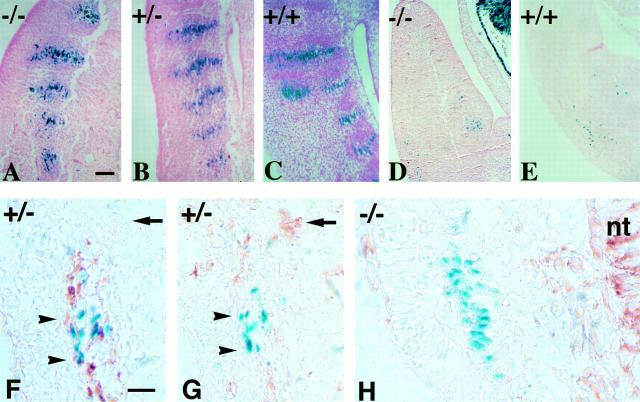
Figure 2.
Myotome differentiation and migration of myogenic cells from somites into limbs of 11-d.p.c embryos. Sagittal sections at the somitic level of Des −/− (A), Des +/− (B), and Des +/+ (C) embryos. Myotomes have the same morphology when revealed by the blue staining. Transversal sections of the limb bud of Des −/− (D), Des +/+ (E) embryos, with the presence in both sections of blue mononucleated myogenic cells, which have migrated but not yet fused. Analysis of vimentin and desmin expression during embryonic development in myogenic cells were performed with immunoperoxidase reactions on cryostat sections on 10.5-d.p.c. embryos from Des +/− heterozygous (G), and Des −/− homozygous (H) mice. The nuclear blue staining was used to characterize the myogenic population. In the control, the desmin-positive, mononucleated cells, which are located in the lateral part of the myotome, have blue nuclei. Immunodetection of desmin stained cytoplasm in brown (F). The adjacent section shows same blue cells negative for vimentin (G). The mutant, mononucleated cells located in the lateral part of the Des −/− myotome are labeled with a blue nucleus but are negative for desmin and vimentin (H). The neural tube (nt) is stained with vimentin antibody. Bar, 50 μm.

