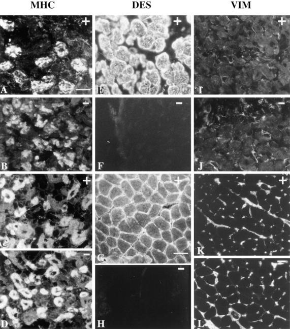
Figure 3.

Detection of myosin isoforms, desmin and vimentin, by immunofluorescence in fetuses and in 1-mo-old mice. Mutant Des −/− (B, D, F, H, J, and L) and control Des +/+ (A, C, E, G, I, and K). (A–D) Characterization of primary and secondary generation muscle fibers at 17.5 d.p.c. Immunofluorescent staining of anterior shoulder muscles on transverse sections of the subscapularis muscle with antibody against slow MHC, which labels primary muscle fibers (A and B) and using an antibody against MHC, which labels secondary muscle fibers (C and D). The same pattern was found in Des +/+ and Des −/− fetuses. (E–H) Detection of desmin using a polyclonal antidesmin antibody showed a typical reactivity in Des +/+ in 17.5-d.p.c. fetuses (E) and 1-mo-old mice (G). No reactivity was found in Des −/− fetus (F) or 1-mo-old mice (H). (I–L) Detection of vimentin using polyclonal anti-vimentin antibody performed on 17.5-d.p.c. fetuses (I and J) and 1-mo-old mice (K and L). The same pattern was found in Des +/+ and Des −/− mice. No vimentin reactivity was found inside the myofibers. The vimentin reactivity found around the myofibers corresponded to connective tissue forming the endomysium and the mesenchymal cells of vessels. Bars: (A–F) 25 μm; (I and J) 25 μm; (G, H, K, and L) 50 μm.
