Figure 5.

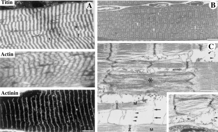
Ultrastructural and immunological characterization of sarcomeres in soleus muscle of Des −/− mice. (A and B) Region of soleus of 8-wk-old Des−/− mice showing sarcomere alignment that is relatively normal or with splitting of the myofibril. (C) In region of soleus of 2-wk-old Des −/− mice with focal alterations. (A) Ultrathin sections stained with antibodies against titin, actin, or α-actinin demonstrate the typical regular striated pattern. However, certain irregularities were observed in the organization of the myofibers that were more easily visualized in the electron microscope. (B) A splitting of the myofibrils can be seen (arrowheads). This splitting is also clearly demonstrated in the ultrathin sections stained with the antibody against titin in A where it can be seen that the Z bands are frequently not in register (arrowheads). (C) Ultrastructure of myofibrillar alterations in the soleus muscle as demonstrated by transmission electron microscopy. On longitudinal sections, filamentous material (arrowheads, top right and bottom center) interlinks the Z disks of one myofibril to the M band region of another myofibril. Another link of filamentous material (arrows) is seen between the M band region of the lower myofibril and the center of two sarcomeres that show Z disk streaming (*). Inset, filamentous material (arrowheads) form myofibril–sarcolemma attachments between the Z disk of a myofibril and dense plaques at the sarcolemma. M, M-line; Z, Z-disk. Bars: (A and B) 5 μm; (C) 1 μm.
