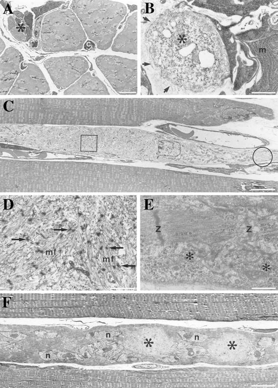
Figure 6.

Transmission electron microscopy of myofibers of soleus from Des −/− 2-wk-old mouse. (A) Cross section of 2-wk soleus showing an area of a normal dense myofibrillar pattern and an area containing several small-size cells. c, capillaries. (B) In higher magnification, the small-size cells are identified as macrophages (m) with well-organized rough endoplasmic reticulum. Activated satellite cells having light cytoplasm with dispersed ribosomes (*). All these cells are enclosed by the same basement membrane (arrows). (C) Longitudinal sections of muscle fiber, one with light cytoplasm, runs in parallel with two other well-organized myofibrils. (D) Higher magnification view of the boxed area in C, showing disorganized myofibrils (mf) and the Z bodies (arrows). (E) Higher magnification view of the encircled area in C, showing an organized sarcomere with Z disks and abundant ribosomes (*). (F) Muscle fiber with areas of light cytoplasm (*) and many large nuclei (n) containing prominent nucleoli run parallel to muscle fibers with well-organized myofibrils. Bars: (A, C, and E) 5 μm; (B and D) 1 μm; (E) 0.5 μm.
