Figure 7.

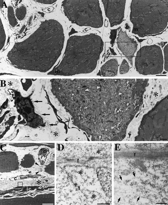
Degeneration and regeneration of soleus from Des −/− 10-wk-old mouse. (A) Transmission electron microscopy of a cross section showing the large variability in fiber diameters seen at 10 wk in the Des −/− soleus. The myofibrillar pattern is well preserved in the largest muscle fibers although abnormal accumulation of mitochondria (m) are present beneath the sarcolemma. Myofibrils are disorganized in some of the small- or intermediate-size fibers. Note that clusters of small fibers occupy the space of a large fiber. The wide interstitial space contains capillaries and cells with slender profiles. (B) Higher magnification of the boxed area in A showing one muscle fiber disorganized myofibrils. An undulating basement membrane (arrows) surrounds interstitial cells with slender processes. Note also the bundles of collagen fibrils (*) in the interstitium. (C) Some muscle fibers show well-organized myofibrils; however, one is divided into three parts, one of which is interrupted by a tendinous junction (arrowheads) after faulty regeneration after fiber damage. Profiles of interstitial cells with slender processes and one cell with a light cytoplasm (*) are seen. (D and E) Higher magnification view of the boxed area in C. In D, strands of myofibrillar material as well as tubules (t) and ribosomes (*) are seen, whereas in E, an array of cytoplasmic filaments are seen beside a myofilamentous strand. Bars: (A and B) 1 μm; (C) 10 μm; (D) 0.5 μm; (E) 0.25 μm.
