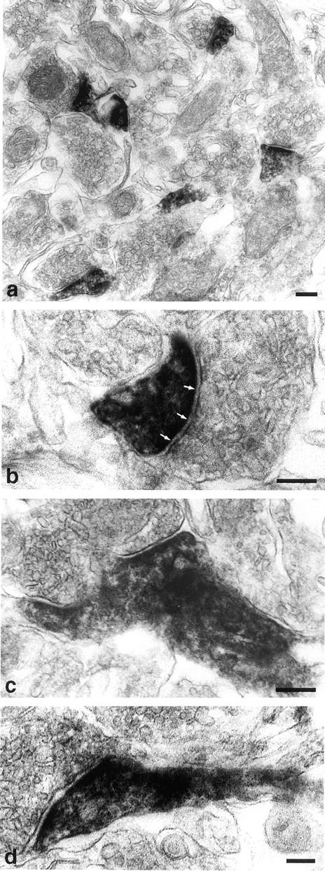
Figure 6.

Immunoelectron microscopic analysis of synaptopodin. The subcellular localization of synaptopodin in the telencephalon was analyzed by preembeding peroxidase labeling. (a) The electron-dense reaction product localizes to the postsynaptic densities and dendritic spines of distinct synapses. (b) At a higher magnification the strongest labeling is found at the PSD (arrows). (c and d) Extension of synaptopodin expression into dendritic shaft. This distribution of synaptopodin in the postsynaptic segment of dendrites corresponds exactly to the distribution of actin at this site. Bars, 0.2 μm.
