
Figure 12.
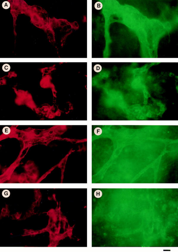
Double immunostaining of normal (+/+) and β1-null (−/−) embryoid bodies for β3 integrin and PECAM (A–D), and αv integrin and PECAM (E–H). Normal and mutant ES cells were differentiated for 20 d, fixed and stained for PECAM (A and C, red) and β3 (B and D, green), or PECAM (E and G, red) and αv (F and H, green), respectively. In normal embryoid bodies large vessels stain for PECAM (A and C) and β3 (B), or αv integrin (D). In β1-null embryoid bodies the vessel diameters are smaller but the staining for PECAM (E and G), β3 (D), and αv (H) is similar, like in normal bodies. Note the high background for αv staining, which is expressed on many PECAM-negative cells in the embryoid bodies. Bar, 15 μm.