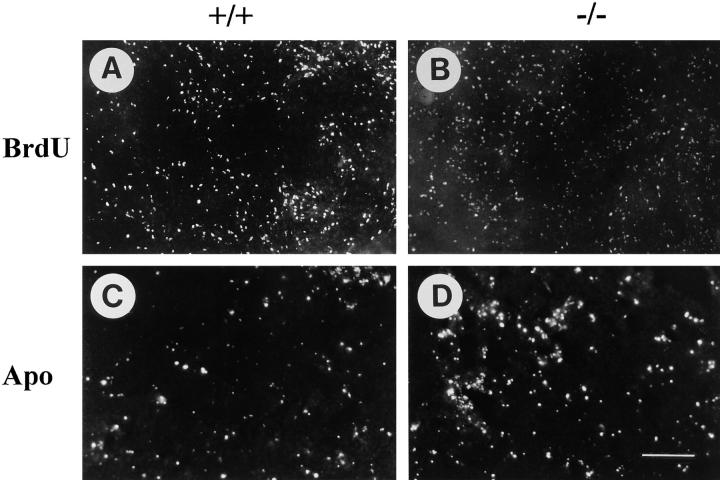
Figure 3.
Analysis of cell proliferation and apoptosis in a normal (+/+) teratoma and a β1-null (−/−) teratoma. ES cells were injected under the skin of the back and incubated for 21 d. 2 h before tumor isolation BrdU was injected intraperitoneally. Incorporated BrdU was detected using specific antibodies labeled with FITC (A and B). Apoptotic cells were identified by staining for the presence of free 3′-hydroxyl groups in tissue sections of +/+ and −/− teratomas (C and D). Bar, 100 μm.

