
Figure 6.
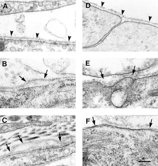
Electronmicroscopy of normal (A and D) and β1-null teratomas and embryoid bodies (B, C, E, and F). Normal cells in teratomas (A) and embryoid bodies (D) are covered by a smooth basement membrane that is strictly located in close vicinity of the cell surface (arrowhead). In β1-null teratomas as well as β1-null embryoid bodies, the basement membranes are partially detached (B and F, arrows), multilayered (C), or show an increased thickness and a loss of typical structure (E). Bar, 250 nm.