Figure 1.
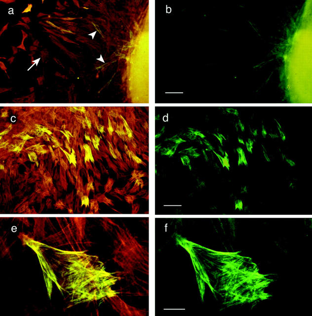
The 1E12 antigen is located intracellularly and codistributes with SMαA in aortic cells cultured from explants on a planar glass substrate. The cells are double labeled with mAbs to SMαA and 1E12. (a, c, and e) Images of cells examined with simultaneous excitation in both fluorescein and rhodamine wavelengths. (b, d, and f) The same fields of cells under fluorescein only excitation, which denotes those cells that are 1E12 positive. (a and b) Low magnification views of aortic cells migrating from explanted tissue. The majority of cells in these fields are labeled with SMαA (arrowhead, red cells). A subset of cells are double labeled with SMαA and 1E12 (arrow, yellow cells). In this field the majority of cells are motile, spindle-shaped cells and, as such, do not show extensive stress fiber arrays, as shown in c and d. (c and d) The explanted aortic cells have migrated onto the planar glass and have formed extensive microfilament (stress fiber) arrays. As above, all the cells in this field were positive for SMαA, while a subset were positive for both SMαA and 1E12. (e and f) A high magnification view of the embryonic aortic cells shows the 1E12 antigen codistributed with actin microfilament bundles. As shown in f, 1E12 labels the entire length of the actin stress fibers in these cells. The faint staining in the other cells is due to optical bleed-through from the rhodamine channel. Bars: (a and b) 50 μm; (c and d) 50 μm; (e and f) 10 μm.

