
Figure 3.
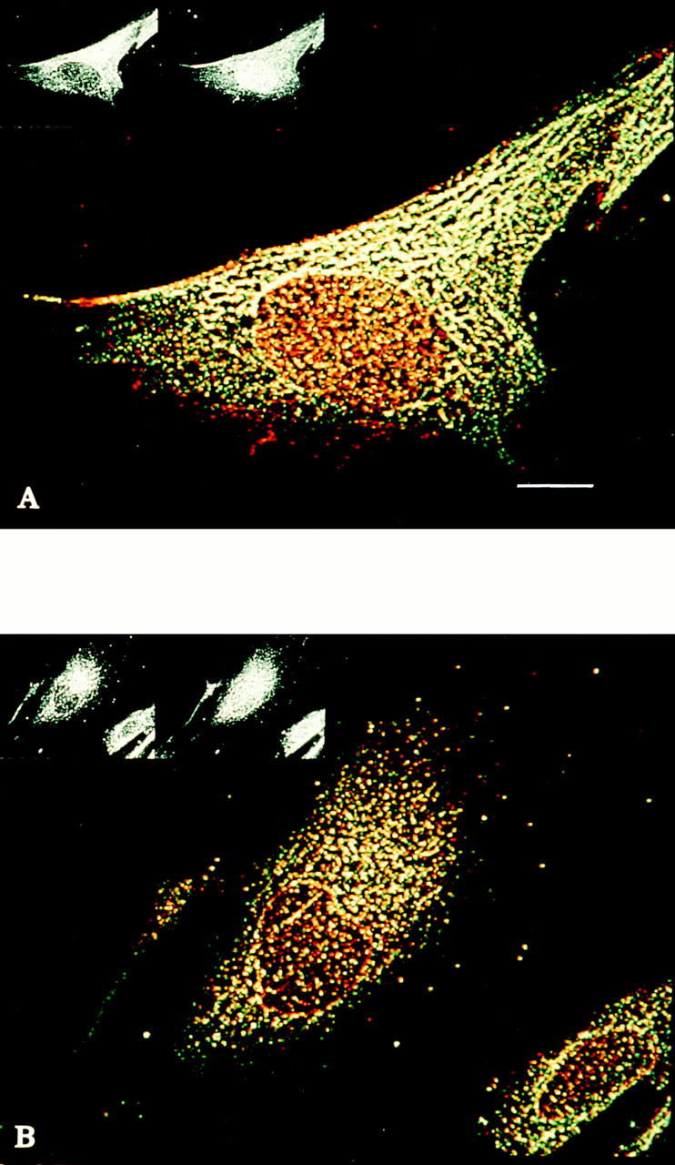
Distribution of ly-hsc73 and lgp120 in serum-deprived and serumsupplemented fibroblasts using a thick optical section. Methanol-fixed fibroblasts were stained with primary antibodies to hsc73 (green) and lgp120 (red) as described in the legend to Fig. 2. The merged color images using an optical section of 4 μm show: (A) serum-deprived fibroblast and (B) serum-supplemented fibroblast. Note that lysosomes appear to fuse, forming a tubular network when cells are serum deprived (A). Bar, 10 μm.