
Figure 3.
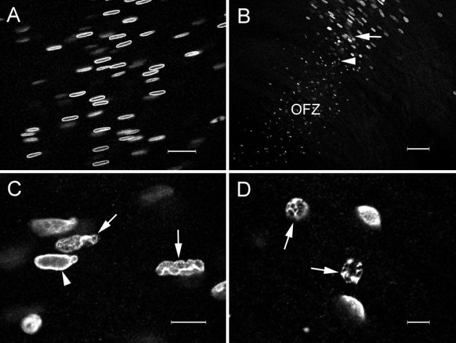
Confocal micrographs of lamin B2 immunofluorescence in an E17 lens slice after high-temperature antigen retrieval (see text for details). (A) Fiber cells in the peripheral cortex. The immunofluorescence is restricted to the lamina of the fiber cell nuclei. (B) Lowmagnification view of cells near the border of the OFZ. Note that even nuclear remnants (arrowhead) are positive for lamin B2 immunofluorescence. (C) High-magnification view of cells immediately adjacent to the OFZ (B, arrow). In many of these cells, the nuclear lamina has become distorted, and the profiles of the nuclei are irregular (arrows), although some nuclei still retain a more normal appearance (arrowhead). (D) High-magnification view of cells immediately within the OFZ (B, arrowhead). At this point in the denucleation process, the fiber nuclei have collapsed into small spherical structures (arrows), and rents are apparent in the nuclear lamina. Bars: (A) 25 μm; (B) 50 μm; (C) 10 μm; (D) 5 μm.