
Figure 6.
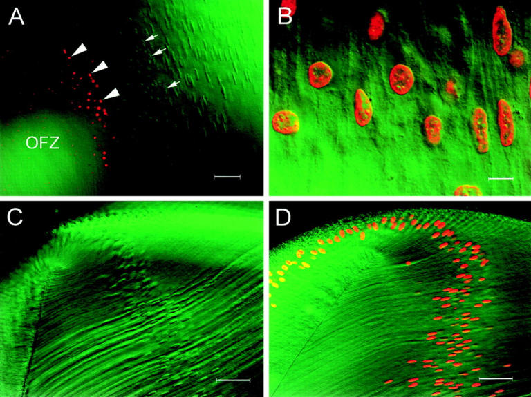
Merged confocal and DIC images of lens slices after TdT labeling with fluorescein-dUTP. The DIC images are shown in green and positively labeled nuclei (containing fragmented DNA) are shown in red. (A) At the border of the OFZ, the nuclei lose their regular shape (arrows) and collapse into condensed structures that are strongly labeled by the TdT assay (arrowheads). Positively labeled debris, resulting presumably from the disintegration of labeled nuclei, extends deep into the OFZ. (B) Cortical fiber cells from a lens slice that was pretreated for 30 min with 50 U/ml DNase I. Note that after DNase I treatment, all nuclei are labeled by the TdT assay and that the labeling is strongest immediately beneath the nuclear membrane. (C) Equatorial region of a lens slice that had been incubated with CIAP before TdT labeling. None of the nuclei are labeled, indicating that the superficial fibers do not contain fragmented DNA with 3′PO4 termini (see text for details). (D) Equatorial region of a lens slice that was treated sequentially with micrococcal nuclease and CIAP before TdT labeling. All the nuclei are labeled, demonstrating the efficacy of the CIAP technique for detecting fragmented DNA with 3′-PO4 termini. Bars: (A) 50 μm; (B) 10 μm; (C) 50 μm; (D) 50 μm.