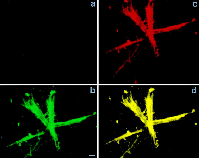
Figure 10.
C-src associates with microtubules generated in vitro. Isolated microtubules were generated from cell lysates and isolated. The microtubules were fixed and stained with both antitubulin and anti–c-src antibodies followed by fluorescent-labeled secondary antibodies, Texas red (for c-src) and FITC (for tubulin), and examined by confocal microscopy. b represents excitation of FITC (tubulin), c represents Texas red (c-src), and d represents both fluorochromes and thus, colocalization of the antibodies. a represents incubation with irrelevant primary murine and rabbit antibodies and FITC and Texas red and excitation of both fluorochromes. Bar, 10 μm.

