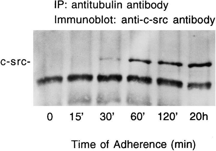
Figure 5.
Time course of c-src/tubulin association. Avian marrow macrophages, maintained in suspension for 2 d, were plated on tissue culture plastic for the indicated times. Cells were lysed, the lysate was immunoprecipitated with antitubulin antibody, and c-src content of the immunoprecipitate was assessed by immunoblotting, as described in Fig. 3.

