
Figure 6.
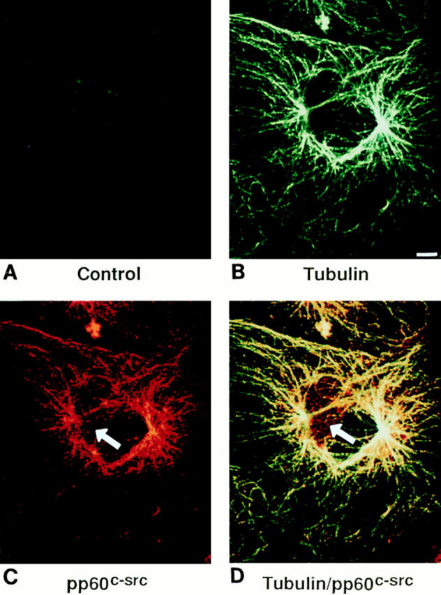
Intracellular localization of c-src and tubulin by immunoconfocal microscopy. Avian marrow macrophages, plated on coverslips, were differentiated into osteoclast-like cells as previously described (1). The cells were fixed, Triton permeabilized, incubated with both antitubulin and anti–c-src antibodies followed by fluorescent-labeled secondary antibodies, Texas red (for src) and FITC (for tubulin), and examined by confocal microscopy. B–D represent differential excitation of the same cell. B represents excitation of FITC (tubulin), and C represents Texas red (c-src). D represents excitation of both fluorochromes and thus, colocalization of the antibodies. The control experiment (A) represents an osteoclast-like cell incubated with irrelevant primary murine and rabbit antibodies and FITC and Texas red and subjected to excitation of both fluorochromes. Bar, 5 μm.