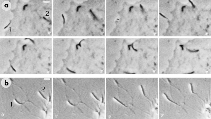
Figure 1.
Video sequences showing Listeria-induced pseudopodium behavior in PtK2 cells (a) and macrophages (b). Time is indicated in min. (a) One bacterium is already in a pseudopodium (1) at time 0′, and the second (2) is in bulk cytoplasm at the head of a comet tail. The pseudopodium (1) gyrates actively for several minutes, and then reenters the main body of the cell at around 5′, forming a typical comet tail (6′ and 7′). The second bacterium begins to form a pseudopodium at 2′, which then exists from 3′ to 5′, after which the bacterium reenters the cell (at 6′), immediately forming a comet (7′). (b) Two pseudopodia (1 and 2) are shown that have already extended away from the macrophage cell body (bottom righthand corner) and continue to move during the sequence, being tethered to the cell by a thin strand. Conditions: (a) phase contrast; (b) Normarski interference contrast. Bars: (a and b) 4.5 μm.

