
Figure 4.
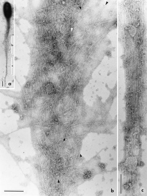
Isolated PtK2 pseudopodium (a) that was well spread in the medial part of the tail. Electron micrographs in b and c show enlargements of the corresponding regions indicated in a. In b the continuity of the axial filaments is particularly evident, when the micrograph is viewed at a grazing angle. The filaments marked with arrowheads, as well as others, can be followed through most of the micrograph. Bars: (a) 1 μm; (b and c) 0.2 μm.