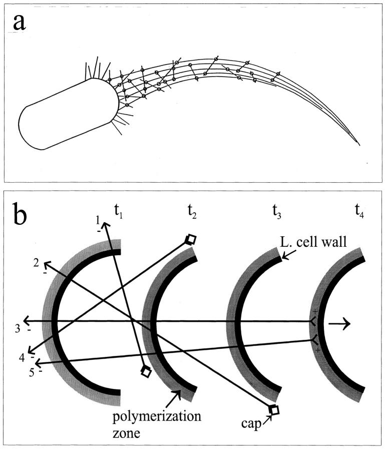
Figure 8.
(a) General organization of actin filaments in pseudopodia. Long filaments provide the axis of the tail, and short filaments cross-link them (circles) into a loose bundle in the region proximal to the bacterium. The comet tails are presumed to have the same structure as the pseudopodia, except that they feature more randomly arranged short filaments. In the more distal parts of the tail, where there are fewer short filaments, the cross-linking between axial filaments predominates (not indicated). (b) Schematic illustration of how the short and long filaments may be generated. The rear end of the bacterium is depicted at different stages of forward movement (arrow) at times t1–t4. The grey, external layer houses components of the polymerization machinery that nucleates actin filaments and feeds their barbed (+) ends with actin monomers. When a filament (+) end lies outside this polymerization zone, it is capped by host cell capping factors (squares). For clarity, only a few actin filaments (straight lines) are shown. Arrowhead configurations indicate the pointed (−) and barbed (+) ends of the filaments. The lengths of a filament will be determined by the position on the rear of the bacterium at which it becomes nucleated and the orientation to the membrane adopted at that time (we assume this to be variable), since this will define how long the filament can reside in the polymerization zone as the bacterium moves forward. The ends of filaments 1 and 4 fall out of the influence of the polymerization zone at t2, after they become tangential to its outer surface. They will be short and obliquely oriented to the tail axis. For filament 2, polymerization ceases at t3. By the same token, more axially oriented filaments (3 and 5) will maintain their plus ends in the polymerization zone for a more extended period and thus become correspondingly longer. L. cell wall, Listeria cell wall.

