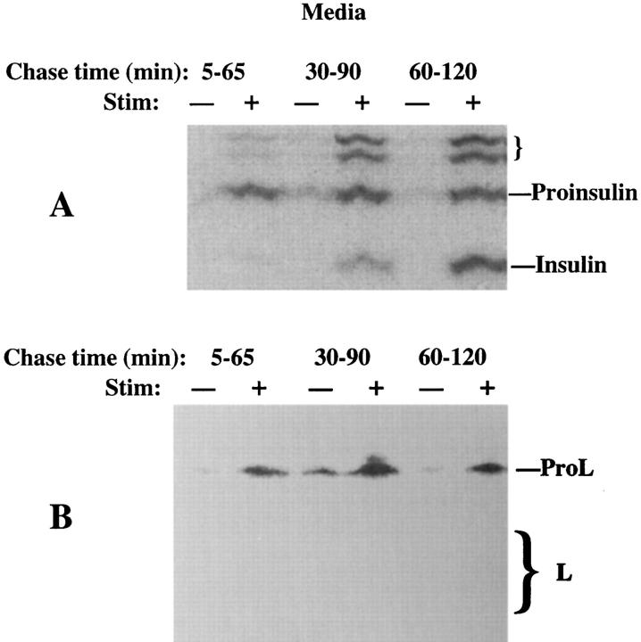
Figure 2.
Stimulus-dependent release from INS cells of peptides immunoprecipitable with antiinsulin (A) or anti–cathepsin L (B). The cells were pulse labeled and chased as in Fig. 1, except that the stimulated (+) or unstimulated (−) medium collections began at 5 min of chase, were 60 min in duration, and were terminated at 2 h of chase. Only the medium is shown. While stimulated secretion of ProL is seen at all chase times, note the progression of processing from proinsulin to insulin in the regulated secretory pathway. Measurement of stimulus-dependent secretion during a 1-h period for L-containing peptides (∼16%) was in a similar range to that of insulin-containing peptides (∼21%) in this experiment. The positions of proinsulin, insulin, presumptive proinsulin conversion intermediates (small bracket), ProL, and bands comprising mature cathepsin L (M r ∼32,000 and ∼27,000, respectively, large bracket) are shown.

