
Figure 10.
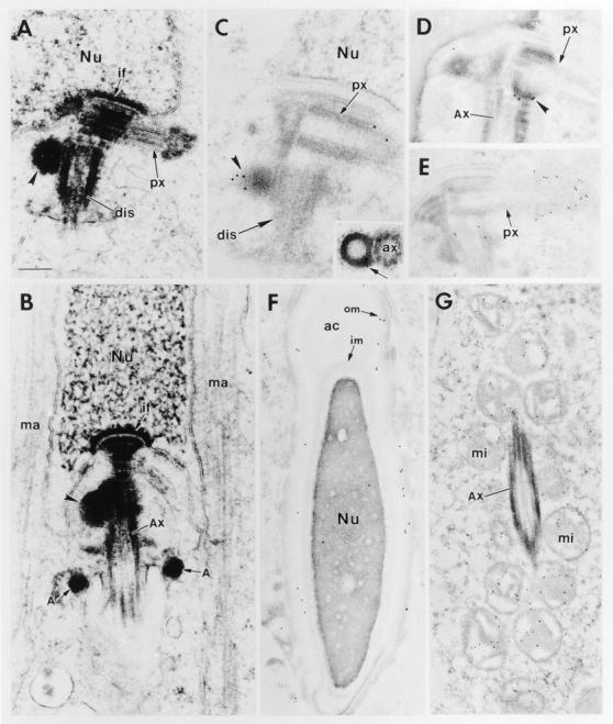
Electron and immunoelectron microscopy of mouse spermatids. Mouse testes from adult animals were fixed and processed for either regular or immunoelectron microscopy as described in the Materials and Methods. For immunoelectron microscopy, we used either αBSN (UC56) or anti–γ-tubulin (Shu and Joshi, 1995) antibodies. (A and B) Negatively stained sections of spermatid centrioles. The tip of the proximal centriole (px) is always associated with the nuclear envelope through implantation foci (if). Its free tip is always encased by a cloud of pericentriolar material; the distal centriole (dis) is always the site of 9 + 2 axoneme assembly and most sections revealed an attached appendage(s) or satellite structure(s) (arrowhead) located near the junction of the two centrioles and often in close proximity to the nuclear envelope. The manchette (ma) of microtubules that surrounds the lower hemisphere of the nucleus is formed at the acrosomal cap phase and disappears shortly after nuclear elongation and flagellar assembly (B). (C and D) Immunogold labeling of centrioles with αBSN. Note that the appendages of the distal centrioles (arrowheads) labeled specifically with the antibody. Inset shows a cross section of an appendage, revealing a hollow center to the structure. (E) Immunogold labeling of centrioles with anti–γ-tubulin. Note that this antibody heavily labeled the pericentriolar cloud at the tip of the proximal centriole and, to a lesser extent, showed some labeling near the distal appendages. (F) αBSN immunogold labeling of the acrosomal cap (ac) of a late-stage spermatid. Note that most of the labeling is concentrated near the outer membrane (om) of the cap rather than the inner membrane (im) or acrosomal space. Note the elongated nucleus, characteristic of nearly mature sperm. (G) Immunogold labeling of the sheath of mitochondria (mi) in the middle piece surrounding the axoneme (Ax). Additional abbreviations: Nu, nucleus; A, annulus. Bar: (A–D) 0.2 μm; (E and F) 0.3 μm; (G) 0.4 μm.