
Figure 4.
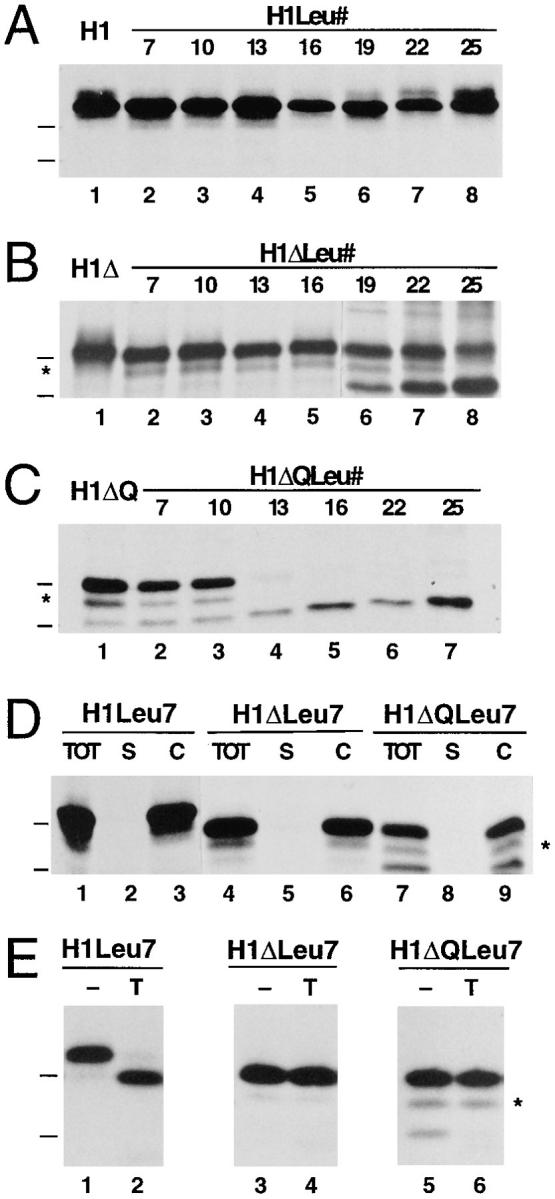
Effect of different hydrophobic domains on membrane insertion of H1, H1Δ, and H1ΔQ. The constructs H1 (A), H1Δ (B), and H1ΔQ (C) with the wild-type transmembrane domain of H1 (lane 1) or with hydrophobic segments consisting of 7–25 leucine residues (lanes 2–8) were expressed in COS-7 cells, labeled, immunoprecipitated, and analyzed by gel electrophoresis and fluorography. Membrane integration assessed by saponin extraction (D) and protease sensitivity (E) is shown for the constructs with the shortest hydrophobic segments of 7 leucines (see legend to Fig. 2). The position of the marker proteins of 29 and 35 kD are indicated.