
Figure 2.
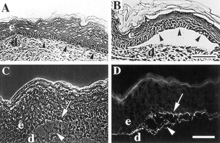
α3-null mice form skin blisters. (A and B) Frozen skin sections from wildtype (A) or α3-null (B) mice were stained with hematoxylin and eosin and the epidermal-dermal junctions were compared. Arrowheads point to basal keratinocytes of the epidermis. (C and D) A frozen skin section from an α3null mouse showing a blister viewed by phase contrast (C) or stained by immunofluorescence with an antiserum against laminin-5 (D). Arrowheads and arrows point to areas of laminin-5 staining at the dermal and epidermal sides of the blister, respectively. e, epidermis; d, dermis. Bars, 50 μm.