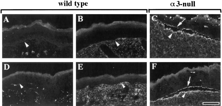
Figure 5.
Fibronectin distributes to both epidermal and dermal sides of blisters in α3null skin. Frozen sections from wild-type (A, B, D, and E) or α3-null (C and F) skin were stained with an antiserum against fibronectin (B and C) or the preimmune serum (A), or with antiserum specific for the EIIIB segment of fibronectin (D–F). Recognition of fibronectin by anti-EIIIB requires treatment with N-glycanase (E and F), as described previously (Peters and Hynes, 1996); as a control, the section in D was not treated with N-glycanase. (A, B, D, and E) Arrowheads point to the dermal-epidermal junction. (C and F) Arrowheads and arrows point to the dermal and epidermal sides of blisters, respectively. Bar, 50 μm.

