Figure 7.
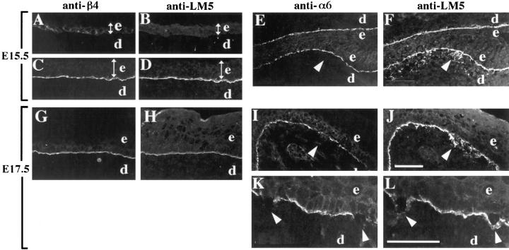
Distributions of α6β4 and laminin-5 in the developing skin of normal and α3-null embryos. Frozen sections from mouse embryonic skin at days E15.5 (A–F) or E17.5 (G–L) of development were stained by double-label immunofluorescence with either monoclonal antibody 346-11A against the β4 integrin subunit (A, C, and G) or GoH3 monoclonal antibody against the α6 integrin subunit (E, I, and K), and anti–laminin-5 serum (B, D, H, and F, J, L, respectively). Control sections were from wild-type embryos (A–D) or heterozygous embryos (G and H). In wild-type E15.5 embryos, α6β4 and laminin-5 codistributed to the basement membrane zone in more stratified regions (C and D), but not in less stratified regions (A and B); the width of the epidermis in each panel is indicated by a double-headed arrow. In α3-null embryos at E15.5 (E and F) and E17.5 (I and J), arrowheads point to areas of laminin-5 staining in areas of disorganized basement membrane, below the α6-positive basal keratinocytes; the skin in E and F is folded back on itself. (K and L) Higher magnification of α3-null skin at E17.5 showing α6-negative, basal keratinocytes that have separated from the laminin-5 positive basement membrane, marked by arrowheads. e, epidermis; d, dermis. Bars: (shown in J for A–J) 50 μm; and (in L for K and L) 50 μm.

