Figure 2.
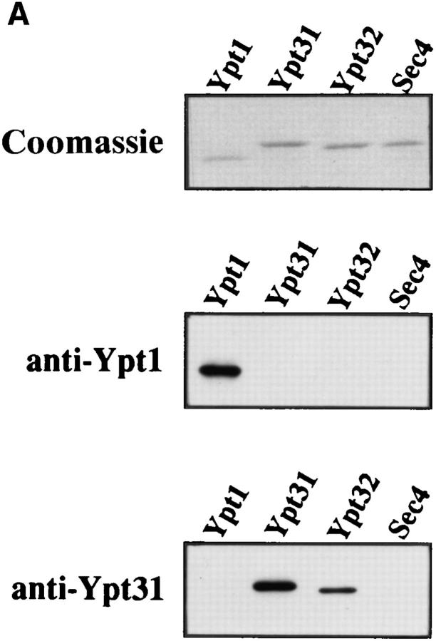
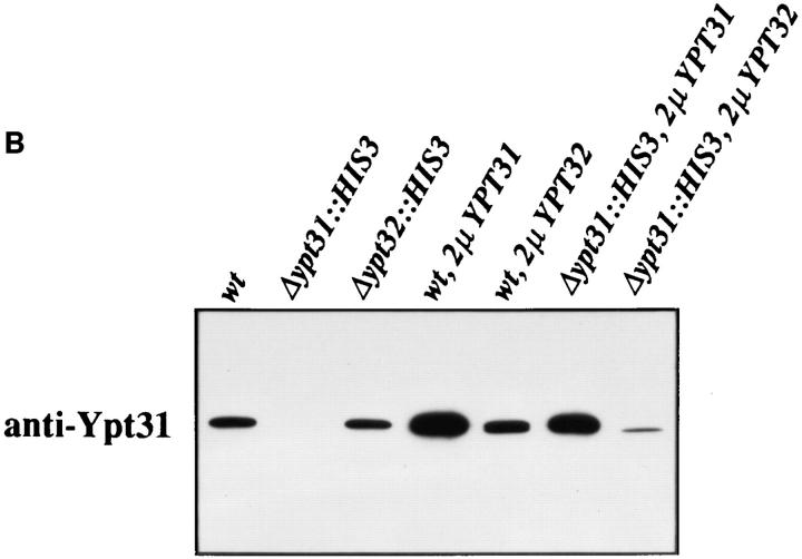
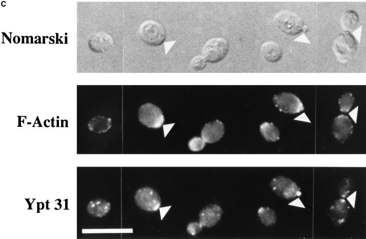
Ypt31 protein is found at sites of polarized cell growth. (A) Characterization of anti-Ypt31 antibodies. Antibodies raised against Ypt31p react with Ypt31p, and to a lesser degree with Ypt32p, but not Ypt1 and Sec4 proteins. Bacterially purified Ypt1, Ypt31, Ypt32, and Sec4 proteins were analyzed by SDS-PAGE followed by either Coomassie staining (1 μg protein in each lane) or Western blotting (5 ng protein in each lane) using anti-Ypt1 or anti-Ypt31 antibodies. (B) Detection of Ypt31 and Ypt32 proteins in yeast cell extracts: Ypt31p is present in excess of Ypt32p in wild-type yeast cells. Wild-type (NSY128) or deletion strains (Δypt31, NSY 290 and Δypt32, NSY296) transformed with the indicated plasmids were grown in synthetic medium maintaining selection for deletions and plasmids present. Cells were lysed by the addition of 2× Laemmli buffer and boiling for 5 min. 0.5 OD600 U of lysate from each strain was then subjected to immunoblot analysis using affinity-purified anti-Ypt31 antibodies. (C) Localization of the Ypt31 protein in yeast cells using immunofluorescence microscopy. Diploid yeast cells (JK9-3d) were double stained with rhodamine-phalloidin (to visualize filamentous actin) and with anti-Ypt31 antibodies. Cells were also photographed using Nomarski optics to distinguish budded from unbudded cells (top). Arrowheads indicate polarized Ypt31 staining, which localizes to regions of cell growth in (from left to right) a cell in late G1, a cell in early S phase, and a cell undergoing cytokinesis. Bar, 10 μm.


