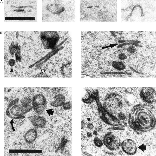
Figure 9.

Details of Golgi structures in wild-type and ypt31-Δ/ypt32-A141D mutant cells. (A) Electron microscopy of wild-type (NSY128) cells. Wild-type Golgi is usually observed as an elongated or cup-shaped single cisternae (Preuss et al., 1992), which are not always continuous (third panel from left) because of fenestration (Rambourg et al., 1993). (B) Enlarged ypt31-Δ/ypt32-A141D mutant (NSY348) Golgi structures. Long arrows indicate an example of stacked cisternae in the mutant strain. Short arrows indicate spherical and tear drop–shaped Berkeley bodies. The tear-shaped structure suggests that Berkeley bodies are derived from individual cup-shaped cisternae that fuse at the rim (see curved arrow for a possible intermediate in this process). Open arrow indicates swelling to 100 nm at the cisternal rim, which may represent an intermediate preceding vesicle formation by membrane fission. Arrowheads indicate rare 100nm vesicles seen in the vicinity of cisternae, for size comparison with structures that look like budding vesicles. Note also the increased length of cisternal profiles in the mutant strain compared to wild-type. Regions of four different cells are shown for each strain. Bars, 0.5 μm.
