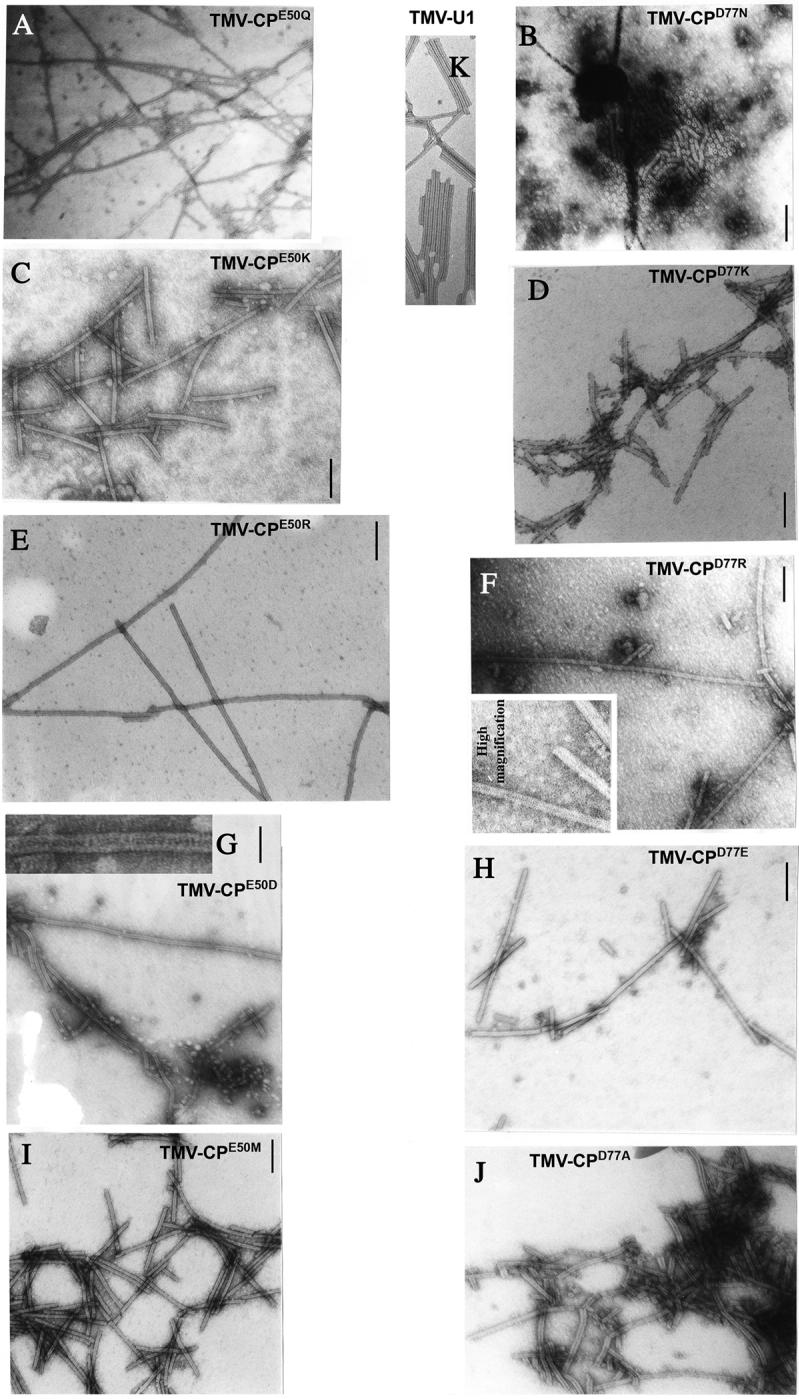
Figure 2.

Electron-microscopic analysis of assembled VLPs from N. tabacum plants infected with TMV mutated at amino acid E50 (TMV-CPE50Q, TMV-CPE50K, TMV-CPE50R, TMVCPE50D, TMV-CPE50M) or at amino acid D77 (TMV-CPD77N, TMV-CPD77K, TMV-CPD77R, TMV-CPD77E, TMV-CPD77A) or w.t. (TMV-U1). The samples were negatively stained and analyzed at a magnification of 39,000. The bar represents 100 nm.
