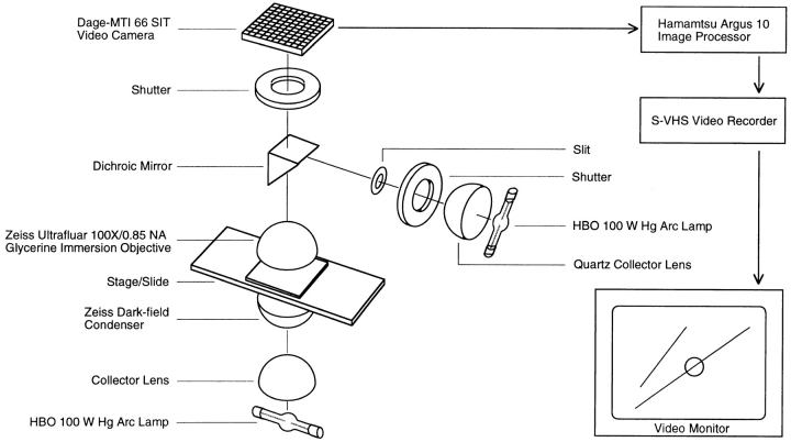
Figure 1.
Schematic diagram of the darkfield UV microbeam apparatus. Self-assembled microtubules were observed by video darkfield microscopy using an HBO 100-W mercury arc lamp, a darkfield condenser (Carl Zeiss, Inc.), a 100×/0.85 NA glycerin immersion objective (Carl Zeiss, Inc.), and a camera (model 66 SIT; Dage-MTI, Inc.). An HBO 100-W mercury arc lamp served as the UV source. A quartz collector lens directed the lamp output through an adjustable diaphragm (the slit) and onto a dichroic mirror, which reflected wavelengths <400 nm to the Ultrafluar objective. The slit image was then projected by the objective onto the specimen image plane. Design details for the integration of the UV microbeam and the darkfield microscope are provided in the text.

