Figure 6.
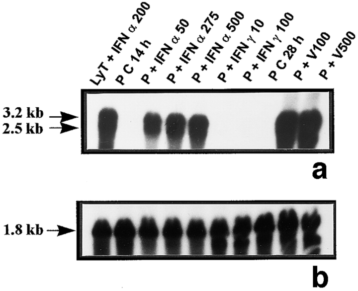
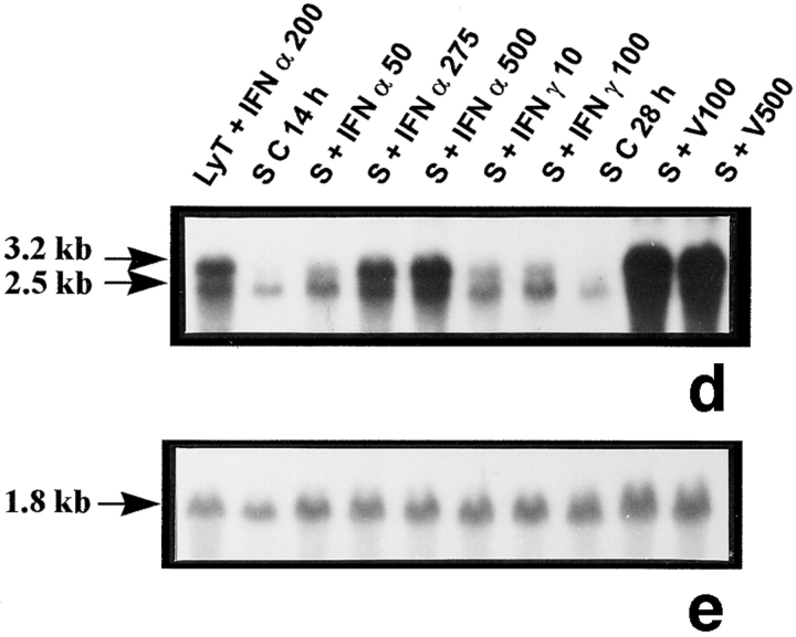
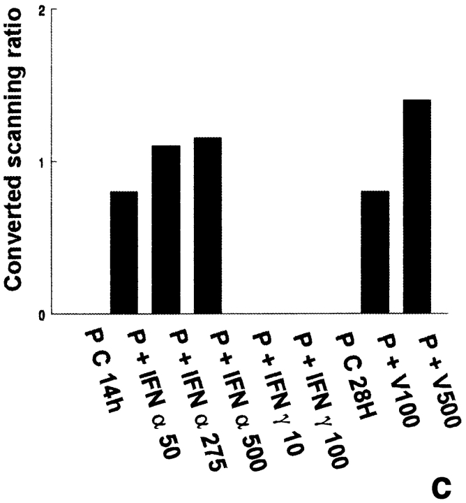
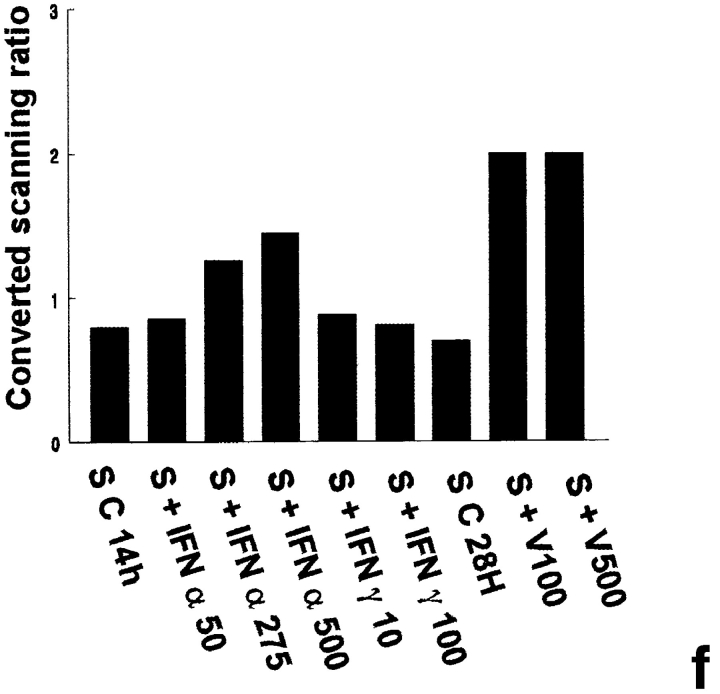
Mx mRNA expression in peritubular and Sertoli cells. Mx mRNA expression was analyzed by Northern blot in peritubular cells (a, P) and in Sertoli cells (d, S) cultured in the presence or in the absence of IFN α (50, 275, and 500 U/ml) or IFN γ (10 and 100 U/ml) for 14 h, or Sendai virus (V100 and V500 U/ml) for 28 h. Hybridization of the blot with the actin probe is shown (b and e). Blots shown are representative of three totally independent culture and Northern blot experiments. mRNA signals were quantified by scanning densitometry and corrected relative to actin signals for both peritubular (c) and Sertoli cells (f).


