
Figure 8.
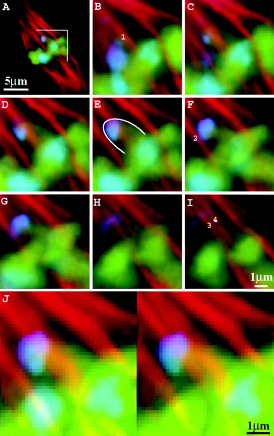
Microtubules interact laterally with neocentromeres. Images are from a heterozygous Ab10 meiocyte (high-knob background) at early metaphase II. (A) Optical section of the entire cell; (B–I) consecutive optical sections at 0.2-μm intervals over the region indicated in A; (J) images in B through I presented as a volume-rendered stereo pair. In B, F, and I, two spindle fibers that contact the knob (1 and 2) and two others that may contact it (3 and 4) are indicated. Chromosomes are shown in green; neocentromeres in blue; microtubules in red. The blue staining in the spindle region of B is derived from knobs that are out of the plane of focus in A. The neocentromere is outlined in E.