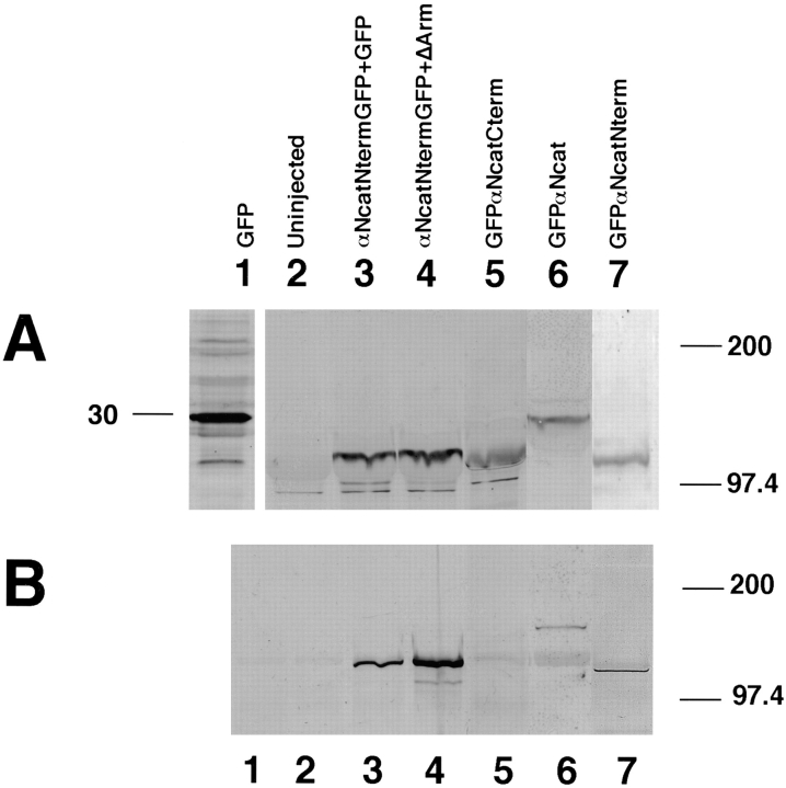
Figure 2.
Expression of α-catenin–GFP fusion proteins in Xenopus embryos and the localization of the β-catenin binding site. (A) Expression of proteins detected by anti-GFP. (B) After immunoprecipitations with anti–β-catenin, associated α-catenin was detected using anti-GFP. In both A and B, the numbering is as follows: lane 1, GFP injected; lane 2, uninjected; lane 3, αNcatNtermGFP + GFP; lane 4, αNcatNtermGFP + ΔArm; lane 5, GFPαNcatCterm; lane 6, GFPαNcat; lane 7, GFPαNcatNterm. Note that GFPαNcatCterm, the protein lacking the NH2 terminus of αN-catenin, does not bind to β-catenin (B, lane 5). Results show that the presence of GFP does not prevent α-catenin binding to β-catenin, provided that the NH2-terminal binding domain is present. In addition, coexpression of ΔArm does not affect expression levels. In each case, 1.5 ng total RNA was injected into one blastomere at the two-cell stage.

