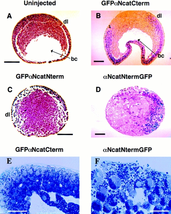
Figure 6.

Histology of gastrulating embryos expressing α-catenin– GFP fusion proteins. Stage 10–11 embryos were fixed in MEMFA and imbedded in paraplast (A and C) or plastic (B, D, E, and F) for sectioning. Arrowheads point to the blastocoel (bc). The abbreviation dl denotes the presumptive dorsal side of the embryos. (A) An uninjected normal embryo. (B) An embryo injected with GFPαNcatCterm mRNA, (C) αNcatNtermGFP, and (D) GFPαNcatNterm. Embryos expressing αNcatNtermGFP completely lack the blastocoel and are disorganized compared to normal embryos. Also noticeable is the lack of integrity of the ectodermal layers in the two mutants. Higher magnification emphasizes the disorganization of cells in an embryo expressing αNcatNtermGFP compared to a normal embryo expressing GFPαNcatCterm. mRNA was injected into the animal pole of both cells of two cell embryos. Bars: (A–D) 200 μm; (E and F) 100 μm.
