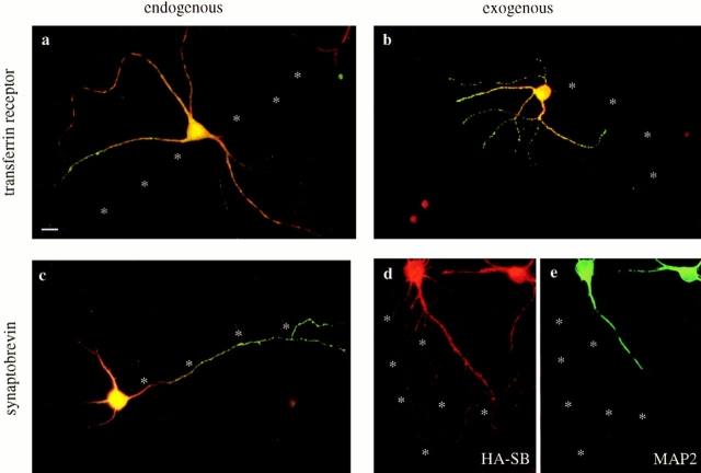
Figure 1.
The distribution of the transferrin receptor and synaptobrevin expressed from defective HSV-1 vectors matches the localization of the endogenous proteins in cultured hippocampal neurons. Neurons cultured for 5–7 d were either fixed immediately, or infected, and then fixed 20 h later. Asterisks indicate the axon. (a) The endogenous transferrin receptor (green) was colocalized with the dendritic marker MAP2 (red) and was strictly excluded from the axon. (b) Human transferrin receptor (green) expressed from a defective HSV-1 vector was also restricted to the dendrites (stained for MAP2 in red). (c) Endogenous synaptobrevin (green) in a control cell infected with the empty HSV-1 vector was concentrated in the distal axon although a small amount of protein colocalized with MAP2 (red) in the dendrites. (d and e) Synaptobrevin with an NH2-terminal HA epitope tag (red) expressed from a defective HSV-1 vector was transported well out into the distal axon (dendrites in e were stained for MAP2 in green). Bar, 20 μm.

