
Figure 3.
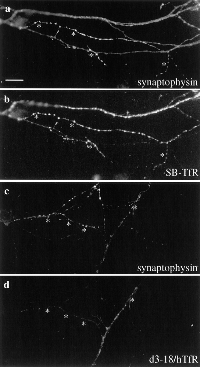
SB-TfR protein in the axon colocalized with the synaptic vesicle marker synaptophysin at synaptic vesicle clusters. Neurons infected with SB-TfR or d3–18/hTfR-defective HSV-1 vectors on day 5–7 were incubated for 20 h and then double-stained with antibodies against hTfR and synaptophysin. (a) Bright spots of synaptophysin staining indicate the location of synaptic vesicle clusters (asterisks). (b) The SB-TfR chimera was colocalized precisely with synaptophysin at many of the puncta. (c) Synaptophysin marks synaptic vesicle clusters in a cell infected with the d3–18/hTfR vector. (d) A mutant transferrin receptor (d3–18/ hTfR) that is not restricted to dendrites was not colocalized precisely with synaptophysin. Bar, 10 μm.