
Figure 8.
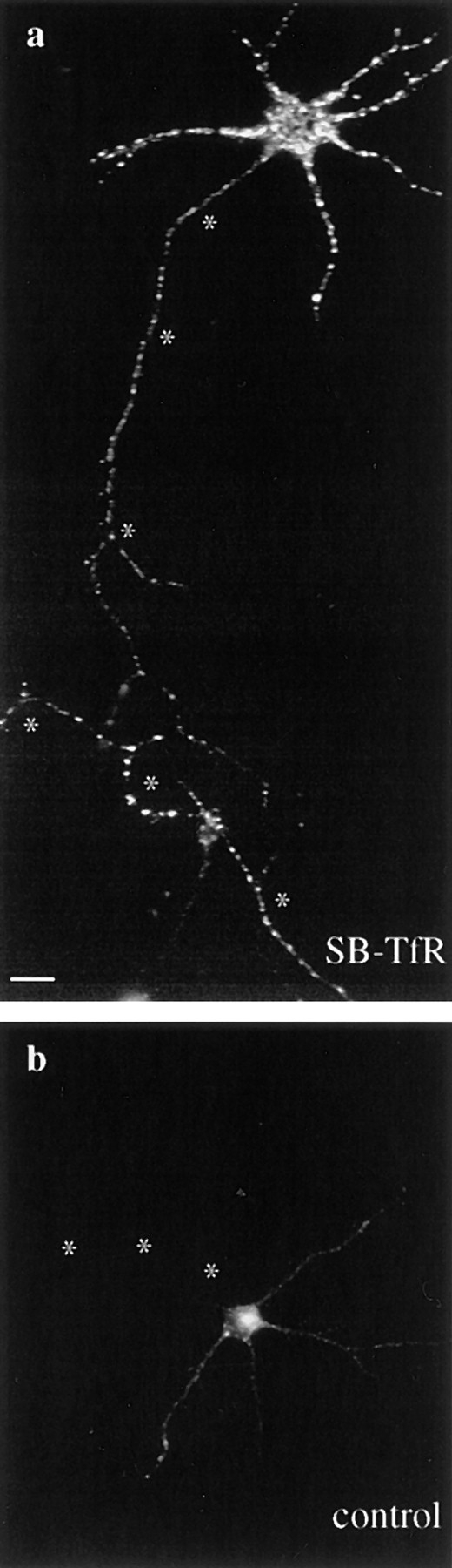
Endocytosis of Cy3-hTf by SB-TfR in axons. Cy3-hTf was added at 37°C to low density cultures of neurons that either had been infected with the SB-TfR vector or were uninfected. After 20 min of labeling, the cells were washed twice, fixed, and then mounted in glycerol for fluorescence microscopy. Asterisks mark the axons. (a) Endocytosed Cy3-hTf labeling of an SB-TfR– infected neuron. Bright puncta were present well out into the long, thin axon. (b) An uninfected neuron showed much less total uptake with no labeling of the axon. Bar, 20 μm.