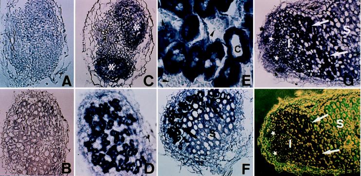
Figure 3.
Localization of MsPG3 expression in root nodule primordia and nodules. In situ hybridization was performed on transversal sections of 4-day-old nodule primordia (A and C), transversal sections of 7-day-old nodules (B, D, and E), and longitudinal sections of 7-day-old (F) and 20-day-old nodules (G and H). Digoxigenin-labeled MsPG3 sense (A and B) and antisense (C–H) probes were used. (E) A higher magnification of the bottom part of D. (H) The same section in G photographed under combination of light and fluorescence microscopy. Asterisks, nodule primordia; stars, meristematic zone; I, invasion zone; S, symbiotic zone; C, infected cells; arrows, interzone II-III; arrow heads, starch grains in noninfected cells.

