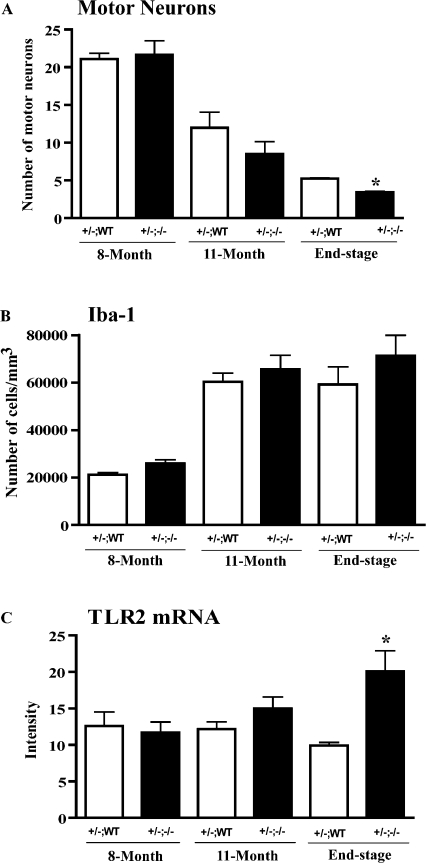
Figure 7.
Number of motor neurons and microglia in the spinal cord of SOD1G37R mice in a MyD88 knockout context. Crossed mice were killed at 8 mo (presymptomatic stage), 11 mo (symptomatic stage), and the end stage. Results represent mean ± the SEM of four mice per group. (A) Motor neurons were counted in the lumbar spinal cord. Note a significant difference between the groups of G37R+/−;MyD88+/+ and G37R+/−;MyD88−/− at the end stage (P < 0.05). (B) Immunohistochemical staining of lumbar spinal cord with the use of microglial cell marker anti–rabbit Iba-1 followed by the incubation with biotinylated secondary antibody. The numbers of microglial cells were estimated and expressed as the number of cells per cubic millimeter. No significant difference was found between the groups of G37R+/−;MyD88+/+ and G37R+/−;MyD88−/− at the presymptomatic, symptomatic, or end stage (P > 0.05). (C) In situ hybridization was performed using antisense probe for TLR2 gene. Quantitative analysis of TLR2 mRNA expression showed a significant difference between the groups G37R+/−;MyD88+/+ and G37R+/−;MyD88−/− at the end-stage (P < 0.05). These data indicate that microglial cells were more activated in G37R+/−;MyD88−/− mice.

