Figure 2.
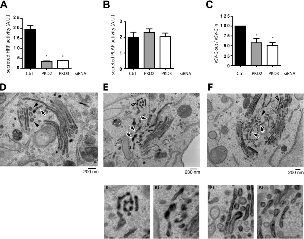
Depletion of PKD2 or PKD3 inhibits secretion of ss-HRP. (A and B) ss-HRP and PLAP cDNAs were cotransfected in HeLa cells depleted or not of PKD2 or PKD3. 20 h after transfection, HRP activity secreted in the medium was measured by chemiluminescence. PLAP activity in cell lysates and secreted into the medium was measured as described in Materials and methods. Bars represent the mean ± SD of HRP (A) or PLAP (B) activity in the medium normalized by PLAP activity in cell lysates/total protein concentration. (C) Depletion of PKD2 or PKD3 inhibits VSV-G transport. tso45VSV-G-GFP plasmid was transfected in HeLa cells depleted or not of PKD2 or PKD3. The levels of VSV-G at the cells surface and inside the cells were determined by FACS analysis. The bars represent the relative ratio of VSV-G at the surface to the total expressed in depleted cells compared with siRNA control transfected cells ± SEM. *, P < 0.01 compared with the control. (D–F) PKD2- and PKD3-depleted HeLa cells and control cells were transfected with ss-HRP and the organization of the Golgi membranes and the localization of HRP monitored by electron microscopy. In control cells (D), the arrow indicates a single tubular profile and arrowheads indicate round profiles (presumably vesicles) in the trans-Golgi region. In contrast, in PKD2- and PKD3-depleted cells (E and F), HRP accumulates in tubular membranes. The arrows indicate multiple tubular profiles at the TGN. The open arrowheads show pearling tubules. The arrowhead in F is a clathrin-coated vesicle revealing the trans side of the Golgi stack.

