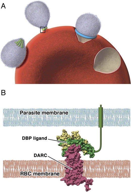
Figure 1. The role of Duffy-Binding Protein in the Invasion of Reticulocytes by P. vivax Merozoites.
(A) Step-wise invasion of reticulocytes involves initial attachment of the merozoite to the cell surface, resulting in deformation of the reticulocyte cell membrane; at this stage DBP is located in the intracellular micronemes (colored green) at the apical end of the merozoite. Reorientation of the merozoite then occurs such that the apical end is in contact with the reticulocyte membrane, and DBP is released for the formation of a tight junction. The tight junction (blue) then moves from the apical to posterior pole as the merozoite invades the cell, propelled by an actin–myosin motor. The reticulocyte membrane is finally re-sealed once invasion is complete. Invasion from initial attachment through to complete entry takes approximately one minute.
(B) A model of the binding of PvDBP to the DARC receptor that occurs during invasion of reticulocytes by merozoites (inset from A). Polymorphic residues are colored yellow; conserved residues are colored green. Antibodies are predicted to bind to a polymorphic region of the DBP that is separate to, but may overlap with, the DARC-binding site.
RBC, red blood cell (reticulocyte)

