Figure 2.
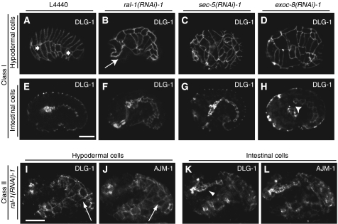
Phenotype of rap-1 mutant embryos carrying the dlg-1∷GFP gene subjected to control, ral-1, sec-5 or exoc-8(RNAi). Phenotypes of rap-1 mutant embryos carrying the dlg-1∷GFP marker (FZ271) that were derived from animals subjected to RNAi for ral-1, sec-5 or exoc-8. DLG-1 indicates dlg-1∷GFP expression, AJM-1 indicates immunofluorescence staining using the MH27 antibody. Genes used for RNAi are indicated above each panel. L4440 is the empty vector control RNAi. (A–H) show class I embryos, (I–L) class II embryos. (A–D) and (I–J) show focal planes to visualize hypodermal cells, (E–H) and (K–L) are focal planes at the level of the gut. The most anterior and posterior visible seam cells are marked with *. In all cases, AJM staining was performed (only shown for class II, subjected to ral-1 (RNAi)), which demonstrated clear colocalization with DLG-1∷GFP). Class I embryos are characterized by halted migration of hypodermal cells as examplified by a ral-1(RNAi) embryo (arrow in B) or misalignment of hypodermal cells as shown here for a sec-5 and exoc-8(RNAi) embryo (C, D). Halted migration can be discerned from early stages of migration by comparison of the developmental stage of the gut. Note that during normal hypodermal cell migration (A, E), the gut has not yet extended along the entire length of the embryos, in contrast to the gut of the ral-1(RNAi) embryo, which has established clear adherens junctions and shows a more mature pharynx (B, F). In some cases, a gap between intestinal and pharyngeal cells is observed (arrowhead H). In class II embryos, hypodermal cells do not envelope the entire embryo (arrow I, J) and a clearly recognizable gut structure is found on the outside of the embryo (arrowhead K). All embryos are oriented such that their pharynx is on the left side. Scale bar: 10 μm.

