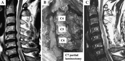
Fig. 1.
Case presentation of selective ELAP. a Preoperative MRI shows three-level stenosis from C4/5–C6/7. The stenosis levels were defined by the disappearance of the subarachnoid space. b Intraoperative photograph. In this three levels stenosis case, the laminae from C4 to C6 laminae were opened in conjunction with upper half laminectomy of C7. c Postoperative MRI

