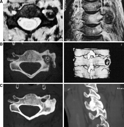
Fig. 1.
a Male 24 years old with OO of the C5 joint pillar in relation to the vertebral artery. b CT scan control 2 months after partial excision through a posterior approach with a mini-access using an operating microscope: residue at approximately midway of the lesion. c CT scan control 4 years after surgical treatment: complete ossification of the lesion. The patient is without symptoms

