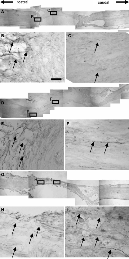
Fig. 2.
Immunohistochemistry for GAP-43 in the MG group (a overview; b, c magnified), BMSC-LacZ group (d overview; e, f magnified), and BMSC-BDNF group (g overview; h, i magnified). b In the MG group, numerous GAP-43-positive fibers were detected at the interface between host spinal cord and graft (arrows). c However, few GAP-43-positive regenerating fibers were detected within the graft (arrows). In contrast, a number of GAP-43-positive regenerating fibers were found at the middle of grafts in the f BMSC-LacZ and i BMSC-BDNF groups (arrows) as well as at the interface between host spinal cord and graft (e BMSC-LacZ, h BMSC-BDNF, arrows). Bars 1 mm (a, d, g) and 30 μm (b, c, e, f, h, i)

