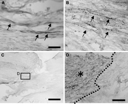
Fig. 4.
Immunohistochemistry for nerve fiber markers in the grafts. a Numerous TH-positive fibers were detected at the middle of the graft in the BMSC-BDNF group (arrows). b Numerous CGRP-positive fibers were detected at the middle of the graft in the BMSC-BDNF group. c, d Only a few 5-HT-positive fibers extended into the graft in BMSC-BDNF rats, although numerous 5-HT-positive fibers were observed at the rostral stump of the host spinalspinal cord [d; higher magnification of box in c; dotted line indicates interface between host spinal cord (asterisk) and graft]. Bars 30 μm (a, b), 500 μm (c), and 50 μm (d)

