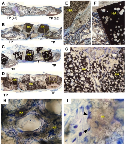
Fig. 4.
Histological sagittal sections of the fusion mass stained with Toluidine Blue O in a rabbit PLF model (a–i). In the Auto (a) group, both bone (b) and cartilage (c) formation can be seen between the L5 and L6 transverse process (TP). In the HA15% (b, e) and the HA50% (c, f) group, newly formed bone (arrows) can be seen between the L5 and L6 transverse process. However, there is only a little direct bone bonding with the HA granules. In these specimens, most surfaces of the HA granules are surrounded by fibrous tissue (f), and bone ingrowth is obtained only in peripheral pores of the implanted HA. In the HA85% (d, g) group, newly formed bone integrates with the HA granules and bone ingrowth is achieved throughout the materials. High magnification photographs in the HA85% (h, i) group show newly formed bone (blue–purple) covers the microporous surface in each macropore of HA (brown) and many osteoblast-like cells (arrowheads) are seen attached to the rough surface of the HA (magnification: a–d ×1, e–g ×5, h ×50, i ×250)

