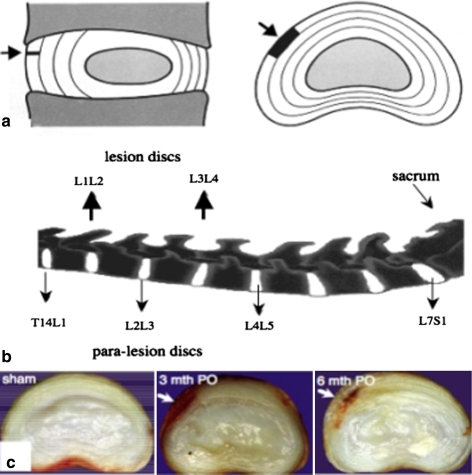
Fig. 1.
a Diagrammatic representation in sagittal and transverse horizontal section of the anatomical site and extent of the anterolateral annular lesion (arrow, dark area). b The L1L2 and L3L4 discs received the lesion depicted in segment a. Para-lesion discs (T14L1, L2L3 and L4L5) were also processed for histology. c Transverse horizontal sections of sham control and lesion discs 3 and 6 month PO depicting the extent of the well defined experimental anterolateral lesion (arrow)

