
Figure 12.
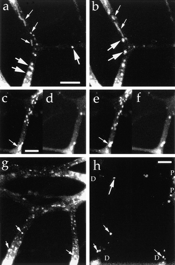
Synaptophysin is transported by the tubulovesicular organelles from the cell body and by the endosomes from the distal axons. Neurons were transfected with adenovirus vector carrying synaptophysin–GFP chimeric DNA. (a and b) Tubular and vesicular organelles with relatively low fluorescence intensity (small arrows) moved anterogradely from the proximal side of the bleached area, whereas large globular vesicles with high fluorescence intensity (large arrows) moved retrogradely from the distal sides. The upper side is proximal to the cell body, and the interval between the frames (a and b) is 20.7 s. (c–f) Double labeling with synaptophysin–GFP (c and e) and FM1-43 (d and f). Tubulovesicular organelles from the proximal side (c and e) were not labeled with FM1-43 (d and f). Signals from retrogradely moving large vesicles were observed using both green (c and e) and red (d and f) filters. Arrowheads indicate the vesicles whose green signal was brighter than the red signal (thus double labeled by both GFP and FM1-43). Interval between the frames in c and d and e and f was 1.65 s. The upper side is proximal to the cell body. (g and h) Double labeling with synaptophysin–GFP and Texas red–dextran (h). P indicates the proximal side of the axons, and D indicates the distal side of the axons. Anterogradely moving tubulovesicular organelles were not labeled with Texas red– dextran, whereas retrogradely moving bright globular vesicles were double labeled with both GFP and Texas red–dextran (small arrows). Note the endosome that was not labeled with GFP (large arrows). Bars, 5 μm.