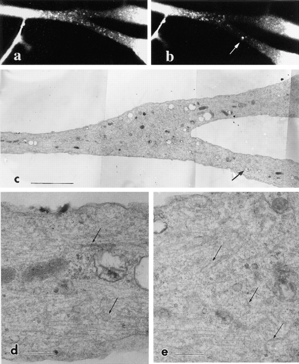
Figure 15.

Correlative electron microscopy of trkA–GFP-transporting vesicles. The axon was photobleached and moving vesicles were observed in the living cell (a). Then the axon was again photobleached and fixed with 2% paraformaldehyde and 0.1% glutaraldehyde. Numerous anterogradely moving vesicles as well as a retrogradely moving large vesicle (arrow) moved into the area before fixation (b). (c) The same area of the axon was prepared for electron microscopy. Arrow in c indicates the corresponding endosome in b. (d and e) Higher magnification of c. Numerous tubules and vesicles were observed (arrows). Bars: (c) 10 μm; (d and e) 1 μm.
